Biomechanical Models of the Human Head
Gertz H.J.1, Hemprich A.2, Hierl Th.2, Kruggel F.3, Meixensberger T.4, Tittgemeyer M.1, Trantakis C.3, Wollny G.1
1 Department of Psychiatry, and 2 Department of Maxillofacial Surgery, University Clinic, Leipzig, 3 University of California, Irvine, 4 Max-Planck-Institute of Cognitive Neuroscience, Leipzig, 5 Department of Neurosurgery, University Clinic, Leipzig
The brain's reaction to mechanical stress (tumor growth, edema), injuries (trauma, hemorrhage), or deforming forces (atrophy, brain growth) was studied by biomechanical forward models: known forces acting on a structural model lead to tissue displacements. Finite element methods (FEM) were employed here that are common in the engineering sciences to model mechanical, electrical or structural properties of systems. Likewise, biomechanical inverse models were studied: observed structural changes with time were analyzed to infer about the underlying forces. A non-linear registration of MRI time-series data leads to a vector field that describes time-dependent changes. A subsequent analysis of this vector field helps in understanding the origin and direction of forces.
 |
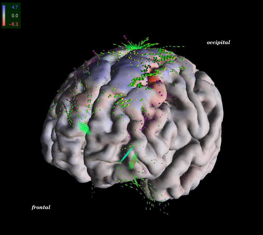
A patient suffering from brain atrophy due to Alzheimer's disease was scanned twice at an interval of 15 months. A displacement field and critical points were computed to visualize the spatial pattern of brain atrophy. A strong repellor in the pre-frontal CSF compartment (see Fig. above) indicates a tissue loss in the vicinity of the frontal pole. Displacement stream lines depict a "flow" of tissue along the midline (as a correlate of a global atrophy). Strong deformations affect the dorsal portions of the first and second frontal gyrus on both sides.
 |
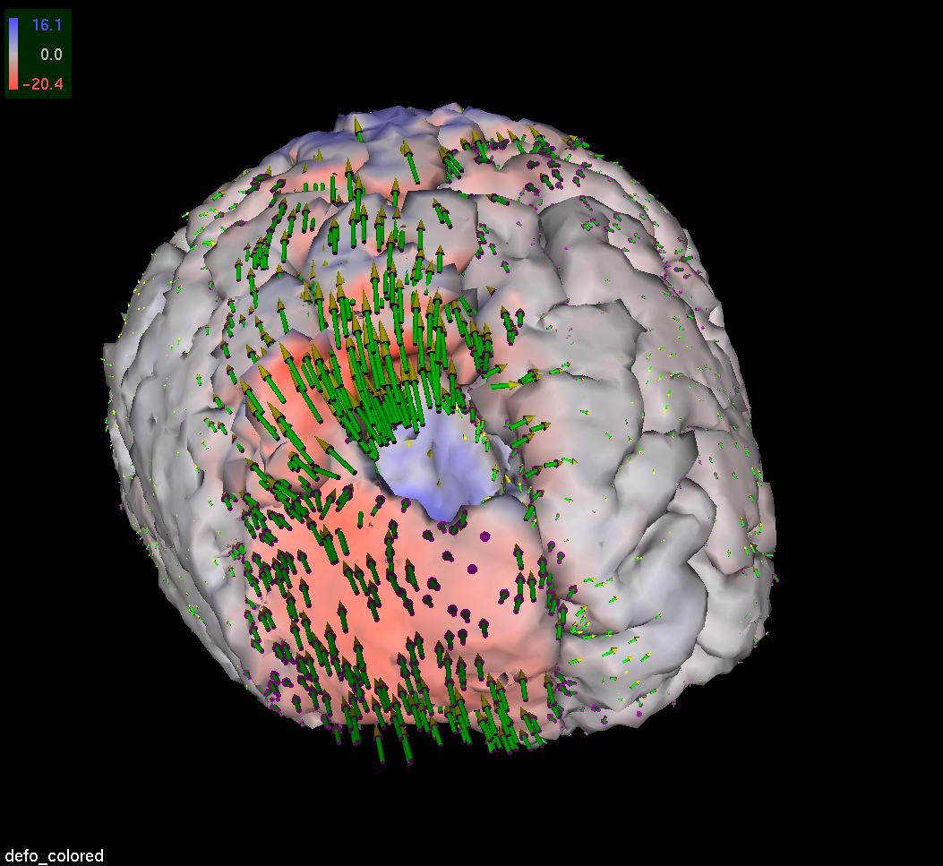
Dramatic structural changes occur during a surgical intervention. MR images were acquired at different timepoints while removing a frontal astrocytoma. Non-linear registration of the timeseries data yields displacement fields describing the brain shift (see Fig. above). Clearly, the shift follows gravity (and increases with consecutive tumor removal), with the major shift at the rim of the tumour. The falx limits the displacement to the ipsilateral side.
 |
 |
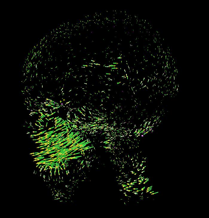
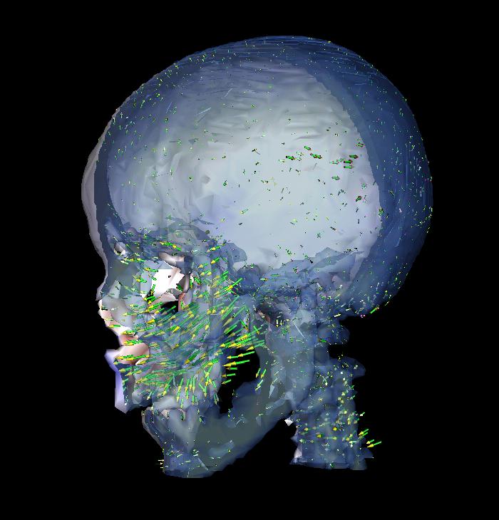
To remedy inborn deformations of the human face, i.e. mainly cleft lip and palate, a metal frame is tightly fixed to the head using screws. After cutting the mid-facial bone along pre-defined lines, this device exerts forces on the bone structures to be relocated. CT datasets were acquired at specific timepoints during the therapy, registered and analyzed for their displacement fields that describe the shift of bone structures in time interval.
Read more...
Hartmann U., Kruggel F. (1998) Transient Analysis of the Biomechanics of the Human Head with a High-Resolution 3D Finite Element Model. Computer Methods in Biomechanics and Biomedical Engineering 2, 49-61.
Hartmann U., Kruggel F. (1999) Modeling Brain Biomechanics. In: Spetzger U., Stiehl H.S., Gilsbach J.M. (eds.) Navigated Brain Surgery, pp. 183-192. Verlag Mainz, Aachen, ISBN 3-89653-553-6.
Hartmann U., Hierl T., Kruggel F., Lonsdale G., Kloeppel R. (2001) Skull mechanics Simulations with the Prototype SimBio Environment. In: Bathe K. (ed.), Proceedings of the First MIT Conference on Computational Solid and Fluid Mechanics (Boston), pp. 243-246. Elsevier, Oxford.
Tittgemeyer M., Wollny G., Kruggel F. (2002) Monitoring Structural Change in the Brain: Application to Neurodegeneration. In: Sporring J., Niessen W., Weickert J. (eds.), Growth and Motion in 3D Medical Images. 3D-Lab University of Copenhagen, ISBN 87-988979-1-8.
