Healthy and Pathological Aging as Revealed by Brain MRI Data
Grunwald M.1, Hensel A.1, Hund-Georgiadis M.2, Kovalev V.A.2, Kruggel F.3, Wolf H.1
1 Department of Psychiatry, University Clinic Leipzig, 2 Max-Planck-Institute of Cognitive Neuroscience, Leipzig, 3 University of California, Irvine
The term "mild cognitive impairment" (MCI) refers to objective cognitive deficits which are not severe enough to fulfill diagnostic criteria for dementia. It has been shown that a certain percentage of MCI patients develop dementia within a few years whereas others remain stable. This study aims at identifying neurobiological parameters that differentiate between the two alternative progression types of MCI. In an ongoing longitudinal study, two-hundred control probands, patients with MCI and demented patients were examined by neuropsychological tests, genetic and peripheral cellular markers, MRI, and EEG. Global measures of brain volume were found to significantly predict changes of cognitive function. Participants with MCI could be distinguished from participants with normal cognition using hippocampal volume measures with an accuracy of 75%.
 |
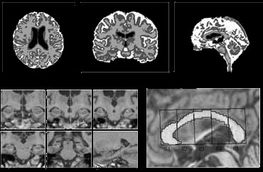
Grey and white matter, internal and external cisterns were automatically determined. Six cross-sections of the hippocampus were segmented manually in the coronal plane on both sides. The corpus callosum was outlined manually in the midsagittal plane.
Read more...
Grunwald M., Busse F., Kruggel F., Riedel-Heller S., Arendt Th., Wolf H., Gertz H.J. (2001) Theta Power Differences in Patients with Mild Cognitive Impairment under Rest Conditions and During Haptic Tasks. Alzheimer Disease and Associated Disorders 16, 40-48.
Grunwald M., Busse F., Hensel A., Kruggel F., Riedel-Heller S., Wolf H., Arendt Th., Gertz H.J. (2001) Correlation Between Cortical Theta Activity and Hippocampal Volumes in Health, mild Cognitive Impairment and mild Dementia. Journal of Clinical Neurophysiology 18, 178-184.
Hensel A., Wolf H., Kruggel F., Riedel-Heller S.G., Nikolaus C., Arendt Th., Gertz H.J. (2002) Morphometry of the Corpus Callosum in Patients with Questionable and Mild Dementia. Journal of Neurology, Neurosurgery and Psychiatry 73, 59-61.
Hund-Georgiadis M., von Cramon D.Y., Kruggel F., Preul C. (2002) Do Questient Arachnoid Cysts Change the Functional Organization in the Brain? A Functional MRI and Morphometric Study. Neurology 59, 1935-1939.
Kovalev V.A., Kruggel F. (2004) A new Method for Quantification of Age-Related Brain Changes. In: 17th International Conference on Pattern Recognition, pp. I-808-811. IEEE Computer Society Press, Piscataway.
Tittgemeyer M., Wollny G., Kruggel F. (2002) Monitoring Structural Change in the Brain: Application to Neurodegeneration. In: Sporring J., Niessen W., Weickert J. (eds.), Growth and Motion in 3D Medical Images. 3D-Lab University of Copenhagen, ISBN 87-988979-1-8.
Wolf H., Grunwald M., Kruggel F., Riedel-Heller S.G., Angerhöfer S., Hojjatoleslami A., Hensel A., Arendt Th., Gertz H.J. (2001) Hippocampal Volume Discriminates Between Normal Cognition, Questionable and mild Dementia in the Elderly. Neurobiology of Aging 22, 177-186.
Wolf H., Kruggel F., Hensel A., Wahlund L.O., Arendt Th., Gertz H.J. (2003) The Relationship Between Head Size and Intracranial Volume in Elderly Subjects. Brain Research 23, 74-80.
Wolf H., Hensel A., Kruggel F., Riedel-Heller S.G., Arendt Th., Wahlund L.O., Gertz H.J. (2004) Structural Correlates of Mild Cognitive Impairment. Neurobiology of Aging 25, 913-924.
