Segmentation of Small Brain Lesions
Descombes X.1, Kruggel F.2
1 INRIA, Sophia Antipolis, 2 University of California, Irvine
As an example of model-based segmentation of pathological structures, consider the detection of multiple small brain lesions from MRI data. A model based on the marked point process framework was designed to detect Virchow-Robin spaces (VRS). These tubular shaped spaces are due to the retraction of the brain parenchyma from its supplying vessels. VRS are described by simple geometrical objects, that are introduced as filters reflecting the VRS geometry in a data term. A prior model includes interactions modeling the clustering property of VRS. An RJMCMC algorithm optimizes the proposed model, obtained by multiplying the prior and the data model. Example results are shown on T1-weighted datasets of elderly subjects. Details are found in reference [Descombes et al. (2003)].
 |
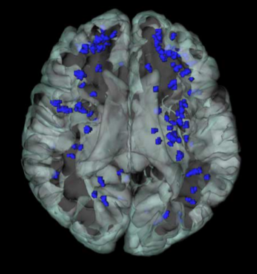
Virchow-Robinson spaces in an example brain. A higher prevalence of VRS is found in the area supplied by the medial and lateral striate arteries (e.g., putamen and pallidum) and in the area supplied by the long penetrating arteries, especially in the frontal white matter compartment.
Read more...
Descombes X., Kruggel F. (2004) An Object-Based Approach for Detecting Small Brain Lesions: Application to Virchow-Robinson Spaces. IEEE Transactions on Medical Imaging 23, 246-255.
Kruggel F., Chalopin C., Kovalev V., Descombes X., Rajapaskse J.C. (2002) Segmentation of Pathological Features in MRI Brain Datasets. In: Gedeon T., Wong P., Halgamuge S., Kasabov N., Nauck D., Fukushima K. (eds.), 9th International Conference on Neural Information Processing (Singapore), pp. 2673-2677.
