EEG-fMRI Coupling
Herrmann C.S.1, Kruggel F.2, Opitz B.1, Wiggins C.J.1
1 Max-Planck-Institute of Cognitive Neuroscience, Leipzig, 2 University of California, Irvine
Measurable correlates of neuronal activation in the brain are electromagnetic fields (by event-related potentials, ERP) and the hemodynamic response (by fMRI) of the vascular system. The first effect is a direct consequence of the electrical activity of neurons, and thus features the same millisecond time scale as the underlying cognitive process, while the second is only indirectly linked to the energy consumption of the neuronal population, and thus takes place on a time scale which is in the order of seconds. Technically, due to the limited number of sensors, the localization of an activation by ERP source analysis is hard to achieve. Here, fMRI features a much easier access to a high spatial resolution. It is obvious that a combination of both techniques is a very attractive aim in neuroscience. However, recording ERPs during fMRI scanning reveals a number of delicate technical problems, requiring a careful experimental set-up and post-hoc signal correction procedures.
 |
 |
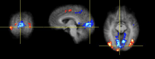
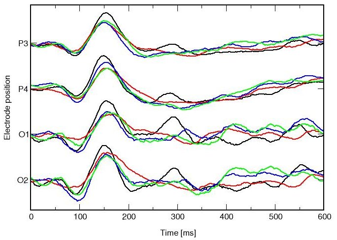
Regions activated by the visual stimulus as detected by functional MRI (top) and ERPs recorded at selected electrode positions (below: black: Kanizsa square, red: Kanizsa triangle, blue: non-Kanizsa square, green: non-Kanizsa triangle).
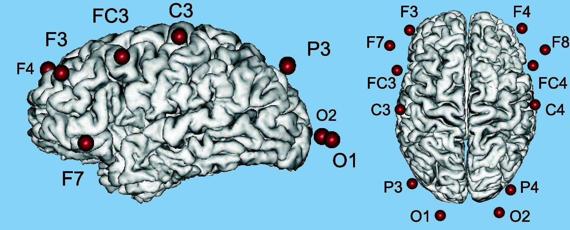
Example results are shown for a well-studied visual oddball paradigm in which Kanizsa and non-Kanizsa figures were used as stimulus material: figures consisted of either three or four inducer disks which we will consider the shape feature and either constituted an illusory figure or not. A total of 900 trials were presented within 45 min. EEG was recorded using 18 electrode in a standard 10/20 set-up while fMRI scanning using an EPI protocol. Images were acquired during a temporal window of 270 ms, leaving between 1730 and 3230 ms for EEG acquisition. FMRI data were analysed using conventional regression statistics and by a adaptation of a non-linear model to the hemodynamic response in the time-series. The same ordering of the amplitude vs. experimental conditions was found for the visual N170 potential and the hemodynamic response of secondary visual cortices. Experimental experience and collected results demonstrate the feasibility of conducting combined EEG-fMRI studies using cognitive tasks [Kruggel et al., 2001].
Read more...
Kruggel F., Herrmann C.S. (2002) Recording of Evoked Potentials During Functional MRI. In: Sommer F. (ed.) Exploratory Analysis and Data Modeling in Functional Neuroscience, pp. 209-228. MIT Press, Cambridge.
Kruggel F., Herrmann C.S., Wiggins C.J., von Cramon D.Y. (2001) Hemodynamic and Electroencephalographic Responses to Illusionary Figures: Recording of the Evoked Potentials During Functional MRI. NeuroImage 14, 1327-1336.
Kruggel F., Herrmann C.S., Wiggins C.J., von Cramon D.Y. (2001) Recording of the Evoked Potentials During Functional MRI: Applications in Cognitive Neuroscience. In: Gedeon T., Wong P., Halgamuge S., Kasabov N., Nauck D., Fukushima K. (eds.), 8thInternational Conference on Neural Information Processing (Shanghai), pp. 77-81.
Kruggel F., Wiggins C.J., Herrmann C.S., von Cramon D.Y. (2000) Recording of the Event-Related Potentials During Functional MRI at 3.0 Tesla Field Strength. Magnetic Resonance in Medicine 44, 277-282.
Opitz B., Mecklinger A., von Cramon D.Y., Kruggel F. (1999) Combining Electrophysiological and Hemodynamic Measures of the Auditory Oddball. Psychophysiology 36, 142-147.

